
عینک eyewear

درباره سایت
آخرین مطالب
- Phantom Vision / Phantom Eye Syndrome
- Vision Stamina and Concussion
- Retinal Tear
- A Theory of Frozen Light
- ? What is Corneal Densitometry (CD)
- ? What are green lenses good for
- the Pars Plana
- Blind Spot Test
- Residual Astigmatism
- Transient Light Sensitivity Syndrome (TLSS)
- health problems associated with computer use مشکلات سلامتی مرتبط با استفاده از کامپیوتر
- decentered ablation correction
- night vision problems after lasik prk epi lasik surgery مشکلات رانندگان لیزیک شده در شب
- eye care after birth مراقبت چشمی پس از تولد
- blue light and blue light control lenses عدسی های کنترل کننده نور آبی
امکانات وب
Pars plana refers to a region of the eye that is characterized by clear, smooth, cystoid cavities known as pars plana cysts, which exist between the pigmented and nonpigmented epithelial layers.
The pars plana (also known as orbicularis ciliaris) (Latin: flat portion) is part of the ciliary body in the uvea (or vascular tunic, the middle layer of the three layers that comprise the eye). It is about 4 mm long, located near the junction of the iris and sclera, and is scalloped in appearance.
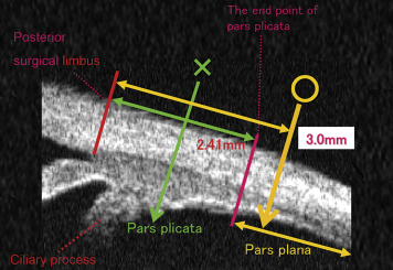
The pars plana constitutes the two-thirds of the ciliary body as the posterior portion. It is a 4 mm wide, smooth surface structure. The pars plana is positioned between the retina and pars plicata and is avascular. Avascular pertains to having little or no blood vessels.
The pars plicata is 2 mm wide and consists of 70 ciliary processes, each approximately 0.5–0.8 mm high and 0.5 mm wide.
The pars plicata (also known as corona ciliaris) (Latin: folded portion) is the folded and most anterior portion of the ciliary body of an eye. The ciliary body is a part of the uvea, one of the three layers that comprise the eye. eResearch by Navid Ajamin -- autumn 2025
What is the difference between pars plana and plicata?
The pars plicata gives rise to the ciliary processes to which the zonules of the lens attach and it surrounds the periphery of the iris. The pars plana has a scalloped posterior border that fits into the scalloped edge of the retina at the ora serrata.
What is the difference between vitrectomy and pars plana?
A vitrectomy performed for diseases of the posterior segment is called a posterior or pars plana vitrectomy. This kind of vitrectomy is performed by a retina specialist. Anterior Vitrectomy: In rare cases, the vitreous gel comes through the pupil into the anterior (front) chamber of the eye.
What are the symptoms of Pars Planitis?
Patients with Pars Planitis usually do not have frank eye pain. What they do notice though is floaters or “stuff” in their vision. In some cases patients with Pars Planitis may go on to develop cataracts and resultant blurry vision.
At pars plana vitrectomy (PPV), changes in ciliary body dimensions with age may affect how sclerotomies are placed so as to avoid iatrogenic damage to the crystalline lens and peripheral retina.
Pars plana vitrectomy is defined as a surgical procedure used to address complicated proliferative diabetic retinopathy and other retinal conditions such as non-clearing vitreous hemorrhage and retinal detachment.
Patients with pars planitis present with minimal symptoms, for example, floaters or blurry vision. In most cases, there is the absence of photophobia and pain. Occasionally patients may present with sudden loss of vision due to retinal detachment or acute vitreous hemorrhage.
Pars plicata refers to the area of the ciliary body that is located between the pars plana and the iris, measuring 2 mm wide. It gives rise to the ciliary processes, which are responsible for attaching the zonules of the lens, and surrounds the periphery of the iris. The non-pigmented epithelium of pars plicata plays a crucial role in the production of aqueous humor, and damage to this area can lead to hypotony.
Reference:
برچسب:
نویسنده: حامد شمس